
The enzymatic degradation of plant polysaccharides is rising as one of the important thing environmental objectives of the early 21st century, impacting on many processes within the textile and detergent industries in addition to biomass conversion to biofuels.
One of the well-known issues with the use of nonstarch (nonfood)-based substrates such because the plant cell wall is that the cellulose fibers are embedded in a community of various polysaccharides, together with xyloglucan, that renders entry tough. There is subsequently growing curiosity within the “accent enzymes,” together with xyloglucanases, that will support biomass degradation by removing of “hemicellulose” polysaccharides.
Here, we report the biochemical characterization of the endo-β-1,4-(xylo)glucan hydrolase from Paenibacillus polymyxa with polymeric, oligomeric, and outlined chromogenic aryl-oligosaccharide substrates. The enzyme shows an uncommon specificity on outlined xyloglucan oligosaccharides, cleaving the XXXG-XXXG repeat into XXX and GXXXG.
Kinetic evaluation on outlined oligosaccharides and on aryl-glycosides means that each the -4 and +1 subsites present discrimination in opposition to xylose-appended glucosides. The three-dimensional constructions of PpXG44 have been solved each in apo-form and as a collection of ligand complexes that map the -Three to -1 and +1 to +5 subsites of the prolonged ligand binding cleft. Complex constructions are in line with partial intolerance of xylosides within the -4′ subsites.
The atypical specificity of PpXG44 could thus discover use in industrial processes involving xyloglucan degradation, reminiscent of biomass conversion, or within the rising thrilling functions of outlined xyloglucans in meals, prescribed drugs, and cellulose fiber modification.
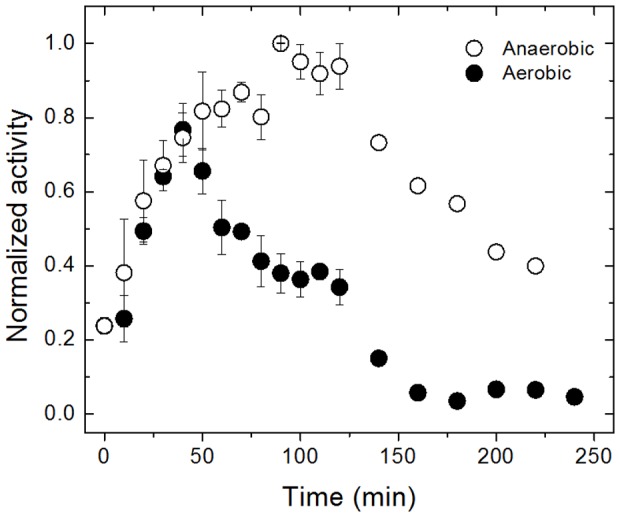
An natural solvent-tolerant lipase from Bacillus sp. pressure 42 was crystallized utilizing the capillary-tube technique. The function of learning this enzyme was with a view to higher perceive its folding and to characterize its properties in natural solvents. By initially fixing its construction within the native state, additional research on protein-solvent interactions might be carried out. X-ray information had been collected at 2.Zero Å decision utilizing an in-house diffractometer.
The estimated crystal dimensions had been 0.09×0.19×0.08 mm. The crystal belonged to the monoclinic house group C2, with unit-cell parameters a=117.41, b=80.85, c=99.44 Å, β=96.40°.
Insolubility of full-length HIV-1 integrase (IN) restricted earlier construction analyses to particular person domains. By introducing 5 level mutations, we engineered a extra soluble IN that allowed us to generate multidomain HIV-1 IN crystals. The first multidomain HIV-1 IN construction is reported. It incorporates the catalytic core and C-terminal domains (residues 52-288). The construction resolved to 2.eight A is a Y-shaped dimer. Within the dimer, the catalytic core domains kind the one dimer interface, and the C-terminal domains are situated 55 A aside.
A 26-aa alpha-helix, alpha6, hyperlinks the C-terminal area to the catalytic core. A kink in a single of the 2 alpha6 helices happens close to a identified proteolytic web site, suggesting that it might act as a versatile elbow to reorient the domains in the course of the integration course of. Two proteins that bind DNA in a sequence-independent method are structurally homologous to the HIV-1 IN C-terminal area, suggesting an analogous protein-DNA interplay during which the IN C-terminal area could serve to bind, bend, and orient viral DNA throughout integration.
A strip of positively charged amino acids contributed by each monomers emerges from every energetic web site of the dimer, suggesting a minimally dimeric platform for binding every viral DNA finish. The crystal construction of the remoted catalytic core area (residues 52-210), independently decided at 1.6-A decision, is similar to the core area throughout the two-domain 52-288 construction.